Monday, 26 August 2013

Cause of Colorectal Cancer May Stem From Mouth Bacteria

Cause of Colorectal Cancer May Stem From Mouth Bacteria

There may be a newly discovered cause of colorectal cancer.
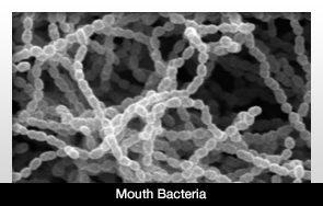
Two new studies indicate that a type of gut bacteria located in the mouth can cause colorectal cancer as a result of influencing the immune response and turning on cancer genes. The research team thinks this information could result in more efficient ways to diagnose, treat and possibly prevent colorectal cancer.
The information from the two studies appears in the August 14 issue of the online journal Cell Host & Microbe.
The gut contains trillions of bacteria, which outnumber the number of cells in our bodies. The microbe communities maintain their health by training the immune system and aiding in the digestion of food. The down side is that they could cause disease.
Previous studies have demonstrated that when there is an imbalance, the bacteria could cause colon cancer.
Of all cancers, colorectal cancer is the second leading cause of death for Americans. The researchers have determined Fusobacteria from the mouth are also plentiful in tissues of colorectal cancer patients.
The first study concluded that Fusobacteria in benign tumors can eventually become cancerous. In mice bred to have a form of colorectal cancer similar to that of humans, the bacteria increased the rate in which tumors formed.
In the second study, it was determined that Fusobacteria utilize a molecule that inhabits the surface of the bacterial cell. It then sticks to and attacks the human colorectal cancer cells.

Stomach Cancer Detection in the Dental Office

Stomach Cancer Detection in the Dental Office

The UCLA School of Dentistry recently received 5 million dollars in funding from the National Institutes for Health to study biological markers in saliva to detect stomach cancer.
There are hopes that this study could advance salivary diagnostics, potentially leading to evidence that saliva can be used to detect other types of cancer, such as pancreatic cancer, breast cancer, and ovarian cancer, as well as diabetes.
The ultimate goal is this: Patients give a saliva sample at the dental office, have it analyzed within minutes, and then are able to find out if they are at risk for stomach cancer.
The research is slated to last five years, and Dr. David Wong is leading the study. As the dentistry school’s associate dean of research, he is considered a pioneer in the field of salivary diagnostics.
Dr. Wong’s team will conduct a prospective study to develop a salivary biomarker panel that would validate stomach cancer detection. The team hopes to capture RNA in saliva samples secreted by stomach cancer cells. This would confirm whether a patient is at risk for stomach cancer.

Study reveals target for drug development for temporomandibular joint disorder (TMJD) - a chronic jaw pain disorder

Study reveals target for drug development for temporomandibular joint disorder (TMJD) - a chronic jaw pain disorder

Main Category: Dentistry
Also Included In: Pain / Anesthetics
Article Date: 05 Aug 2013 - 1:00 PDT

Current ratings for:
Study reveals target for drug development for temporomandibular joint disorder (TMJD) - a chronic jaw pain disorder

Patient / Public:3 stars
3 (1 votes)
Healthcare Prof:not yet rated
Article opinions: 1 posts
Temporomandibular joint disorder (TMJD) is the most common form of oral or facial pain, affecting over 10 million Americans. The chronic disorder can cause severe pain often associated with chewing or biting down, and lacks effective treatments.
In a study in mice, researchers at Duke Medicine identified a protein that is critical to TMJD pain, and could be a promising target for developing treatments for the disorder. Their findings are published in the August issue of the journal PAIN.
Aside from cases related to trauma, little is known about the root cause of TMJD. The researchers focused on TRPV4, an ion channel protein that allows calcium to rapidly enter cells, and its role ininflammation and pain associated with TMJD.
"TRPV4 is widely expressed in sensory neurons found in the trigeminal ganglion, which is responsible for all sensations of the head, face and their associated structures, such as teeth, the tongue and temporomandibular joint," said senior study author Wolfgang Liedtke, M.D., PhD, associate professor of neurology and neurobiology at Duke. "This pattern and the fact that TRPV4 has been found to be involved in response to mechanical stimulation made it a logical target to explore."
The researchers studied both normal mice and mice genetically engineered without the Trpv4 gene (which produces TRPV4 channel protein). They created inflammation in the temporomandibular joints of the mice, and then measured bite force exerted by the mice to assess jaw inflammation and pain, similar to how TMJD pain is gauged in human patients. Given that biting can be painful for those with TMJD, bite force lessens the more it hurts.
The mice without the Trpv4 gene had a smaller reduction in bite force - biting with almost full force - suggesting that they had less pain. In normal mice there was more TRPV4 expressed in trigeminal sensory neurons when inflammation was induced. The increase in TRPV4 corresponded with a greater reduction in bite force.
The researchers also administered a compound to normal mice that blocked TRPV4, and found that inhibiting TRPV4 also led to smaller reductions in bite force, similar to the effects of the mice engineered without the Trpv4 gene.
Surprisingly, the researchers found comparable bone erosion and inflammation in the jaw tissue across all mice, regardless whether the mice had TRPV4 or not.
"Remarkably, the damage is the same but not the pain," Liedtke said. "The mice that had the most TRPV4 appeared to have the most pain, but they all had similar evidence of temporomandibular joint inflammation and bone erosion in the jawbone as a consequence of the inflammation."
The results suggest that TRPV4 and its expression in trigeminal sensory neurons contribute to TMJD pain in mice. Given the lack of effective treatments for this chronic pain disorder, TRPV4 may be an attractive target for developing new therapies.
Stem cells in the gingiva may have important medical applications in the future

Stem cells in the gingiva may have important medical applications in the future

Main Category: Dentistry
Also Included In: Stem Cell Research;  Immune System / Vaccines
Article Date: 07 Aug 2013 - 1:00 PDT

Current ratings for:
Stem cells in the gingiva may have important medical applications in the future

Patient / Public:not yet rated
Healthcare Prof:not yet rated
Stem cells found in mouth tissue can not only become other types of cells but can also relieve inflammatory disease, according to a new Ostrow School of Dentistry of USC study in the Journal of Dental Research.
The cells featured in the study are gingival mesenchymal stem cells (GMSC), which are found in the gingiva, or gum tissue, within the mouth. GMSC, like other stem cells, have the ability to develop into different types of cells as well as affect the immune system.
"Gingiva is very unique in our body," says Professor Songtao Shi, the study's senior author. "It has much less inflammatory reaction and heals much faster when compared to skin."
Previously, the developmental origins and abilities of GMSC hadn't been fully illustrated. This study shows that there are two types of GMSC: those that arise from the mesoderm layer of cells during embryonic development (M-GMSC) and those that come from cranial neural crest cells (N-GMSC). The cranial neural crest cells develop into many important structures of the head and face, and 90 percent of the gingival stem cells were found to be N-GMSC.
The two types of stem cells vary dramatically in their abilities. N-GMSC were not only easier to change into other types of cells, including neural and cartilage-producing cells; they also had much more of a healing effect on inflammatory disease than their counterparts. When the N-GMSC were transplanted into mice with dextrate sulfate sodium-induced colitis - an inflamed condition of the colon - the inflammation was significantly reduced.
The study indicates that the stem cells in the gingiva - obtained via a simple biopsy of the gums - may have important medical applications in the future.
"We will further work on dissecting the details of the gingiva stem cells, especially their notable immunoregulatory property," says first author Xingtian Xu, specialized lab technician at the Ostrow School of Dentistry Center for Craniofacial Molecular Biology.
"Through the study of this unique oral tissue, we want to shed the light on the translational applications for improving skin wound healing and reducing scar formation."

Results from large international clinical trials provide dentists and patients with a new level of confidence in dental implants

Results from large international clinical trials provide dentists and patients with a new level of confidence in dental implants

Main Category: Dentistry
Article Date: 08 Aug 2013 - 2:00 PDT

Current ratings for:
Results from large international clinical trials provide dentists and patients with a new level of confidence in dental implants

Patient / Public:not yet rated
Healthcare Prof:not yet rated
Results from two of the largest international clinical studies performed to date with dental implants have just been published and demonstrate excellent clinical performance. Together, the studies have evaluated more than one thousand Straumann Bone Level implants in Europe, the US and Australia. The scope of these studies is particularly remarkable in view of the fact that most dental implant companies do not conduct clinical trials because they want to cut costs and do not have the capabilities.
The studies are also remarkable in that they both reported very high implant survival rates of more than 98% with practically no bone loss around the implants. The findings are considerably better than values reported in a comprehensive review of previously published studies with other implants[1]. The new studies add to the wealth of strong clinical evidence backing the Straumann dental implant system and thus provide very good reasons for patients and dentists to insist on Straumann implants rather than undocumented alternatives.

The benchmark in clinical research

The first study was a randomized controlled clinical trial (RCT) at 11 clinical centers in Europe, USA and Australia[2]. RCTs are the benchmark in clinical research because they offer the highest level of clinical evidence. However, few are performed with dental implants and very rarely on such a large scale.
This RCT has evaluated 106 patients each treated with one implant and followed for three years. The investigators compared the outcomes of two different approaches - the first involving two surgical steps, in which the implant is covered with gum tissue ('submerged') during healing, and the second involving just a single step, in which part of the implant is left exposed ('transmucosal') thus saving a second surgical operation. The most impressive highlight reported was the fact that only a single implant was lost, yielding 3-year implant-survival rates of 98.1% and 100% for the transmucosal and submerged groups respectively.
Because bone loss around implants has been documented as a common undesirable effect of implant treatment[3], this study looked carefully at bone level changes. It showed that bone level was impressively stable over 3 years after implant placement, with mean decreases of less than 0.7 mm and 0.6 mm in the submerged and transmucosal groups respectively. These values are well below the data presented in previous studies with other implants. An analysis of published data showed that only three implant systems achieved mean marginal bone loss below 1 mm over a period of 5 years[1].

Excellent results also achieved in everyday practice

While RCTs demonstrate that products or treatments work well, they are usually conducted by specialists in selected and strictly controlled populations. This study was performed by dental practices and University clinics that are highly specialized in dental implantology, which raises the question of whether its excellent results can be reproduced in daily dental practice. To answer this, a large study using the same implant was conducted in Europe and the US, in which the dentists had to follow the product guidelines but were able to use the implant as they would in normal daily practice. The strength of this type of investigation, which is known as 'non-interventional study' (NIS), is that it documents real-life situations, in which indications, patients and conditions all vary widely.
In this study[4], a total of 908 implants were evaluated in 538 patients at more than a hundred dental practices in six countries, revealing an implant survival rate of 98.5% after one year (the risk of failure is highest in the first year after implant placement5). Besides the very high survival rates, the bone level remained very stable in the majority of cases. The investigators therefore concluded that treatment with Straumann Bone Level Implants yielded very successful outcomes in 'real life' conditions.

Results impress further when viewed in the context of other published data


The survival rates reported in both studies are higher than those documented in the literature. The most recent analysis of published data on other implants showed an overall implant survival rate of 95.5% one year after implant placement[5], in contrast to the 98.5% achieved in this NIS in daily dental practice conditions.